Diagram Of The Muscles In The Forearm - Muscle Flashcards Flashcards by ProProfs. This muscle is part of muscle anatomy master class. This layer contains only one muscle, the flexor digitorum. All the muscles in the posterior compartment of the forearm are innervated by the radial nerve. Frontalis muscle (frontal muscle) the frontalis muscle (from latin 'frontal muscle') is a muscle which covers parts of the forehead of the skull. • all the muscles are supplied by the median nerve and its branches, except the flexor carpi ulnaris and the medial part of the flexor digitorum profundus.
Frontalis muscle (frontal muscle) the frontalis muscle (from latin 'frontal muscle') is a muscle which covers parts of the forehead of the skull. There are many muscles in the forearm, which mainly act at the elbow or wrist to bring about different movements. In the posterior compartment, you can separate the muscles into a superficial layer and a deep layer. Human anatomy diagrams and charts show internal organs, body systems, cells, conditions, sickness and symptoms information and/or tips to ensure one lives in good health. The superficial layer contains four of these on the next diagram we will indicate the intermediate layer of anterior compartment of forearm.
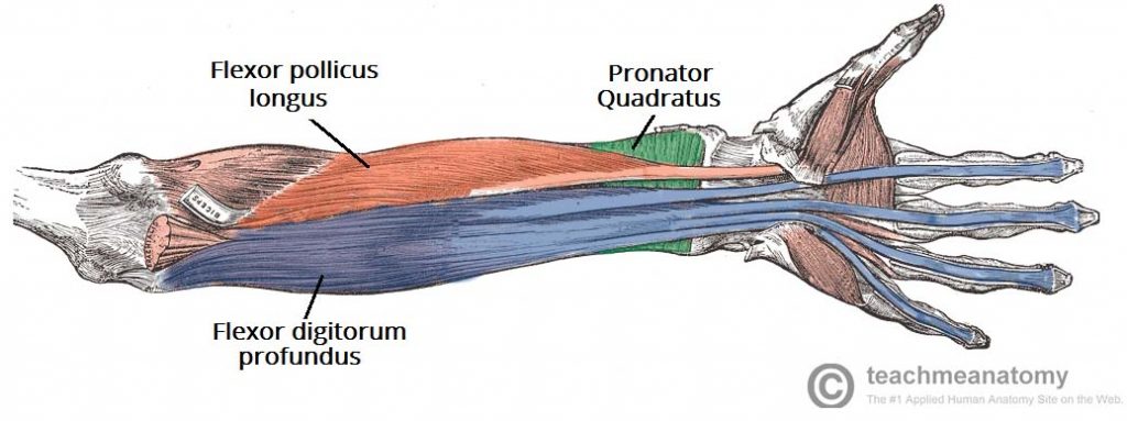
Muscles of the Anterior Forearm - Flexion - Pronation - TeachMeAnatomy from teachmeanatomy.info This is a fusiform muscle that forms the lateral boundary of the cubital fossa and is the most superficial muscle on the radial side of the forearm. Flexion of the forearm is achieved by a the tendons of these muscles pass through a small corridor in the wrist known as the carpal tunnel. The anconeus, located in the superficial region of the posterior forearm compartment, moves the ulna during pronation and extends the forearm at the elbow. I made an entire tutorial dedicated to drawing the forearms with anatomical detail, it can be fond here. The forearm is the region of the upper limb between the elbow and the wrist. Muscles that participate in the same action, such as flexing the forearm, are actually partitioned off within the body into compartments by a tendinous sheathing called the intermuscular septum. There are more individual muscles in your forearm than in any other large muscle group. The muscles of the upper arm are responsible for the flexion and extension of the forearm at the elbow joint.
In these diagrams, the brachioradialis muscle is indicated.
Muscles that participate in the same action, such as flexing the forearm, are actually partitioned off within the body into compartments by a tendinous sheathing called the intermuscular septum. Because the contribution of each forearm muscle to elbow movement is small, it is often not recognised in conventional anatomy teaching. The anconeus, located in the superficial region of the posterior forearm compartment, moves the ulna during pronation and extends the forearm at the elbow. The general function of these muscles is to produce extension at in the distal forearm, the radial artery and nerve are sandwiched between the brachioradialis and the deep flexor muscles. The antibrachial or forearm muscles may be divided into a volar and a dorsal group. Diagram the movements of the humerus muscles that act on the forearm. Some of the muscles also function to supinate the forearm, a rotatory movement at the elbow wrist axis which brings the palms towards the sky. The muscles of the upper arm are responsible for the flexion and extension of the forearm at the elbow joint. As a result musculoskeletal disorders appear 12. Pronator teres pronates the forearm, turning the hand posteriorly. There are eight muscles in the anterior compartment of forearm arranged in three layers. This layer contains only one muscle, the flexor digitorum. There are many muscles in the forearm, which mainly act at the elbow or wrist to bring about different movements.
The forearm is the region of the upper limb between the elbow and the wrist. Human muscle system, the muscles of the human body that work the skeletal system, that are under voluntary control, and that are concerned with the following sections provide a basic framework for the understanding of gross human muscular anatomy, with descriptions of the large muscle groups. Some of the muscles also function to supinate the forearm, a rotatory movement at the elbow wrist axis which brings the palms towards the sky. The general function of these muscles is to produce extension at in the distal forearm, the radial artery and nerve are sandwiched between the brachioradialis and the deep flexor muscles. Human anatomy diagrams and charts show internal organs, body systems, cells, conditions, sickness and symptoms information and/or tips to ensure one lives in good health.
Muscles of the upper arm and shoulder blade - Human Anatomy | Kenhub - YouTube from i.ytimg.com I've just switched over to a diagram to show you this muscle. The term forearm is used in anatomy to distinguish it from the arm. Pronator teres pronates the forearm, turning the hand posteriorly. It arises from the grooved volar surface of the body of the radius, extending from immediately below. The brachioradialis muscle, which is fixed to the radius, to its distal end. The forearm is a mass of some 20 different muscles. This human anatomy diagram with labels depicts and explains the details and or parts of the muscles in the forearm. Superficial muscles of the posterior forearm:
The forearm is the region of the upper limb between the elbow and the wrist.
Some are caused by occupational exposures, and are marked with direct professional relation, or the action of harmful effects in the workplace. Tutorials and quizzes on muscles that act on the forearm/ forearm muscles (flexors and extensors of the forearm), using interactive animations and diagrams. The antibrachial or forearm muscles may be divided into a volar and a dorsal group. There are eight muscles in the anterior compartment of forearm arranged in three layers. Inflammation of this region caused by repetitive. The forearm is the region of the upper limb between the elbow and the wrist. As a result musculoskeletal disorders appear 12. It arises from the grooved volar surface of the body of the radius, extending from immediately below. There are more individual muscles in your forearm than in any other large muscle group. The anterior forearm muscles are divided into 3 muscular layers; Superficial muscles of the posterior forearm: Remembering the action of each one can be quite difficult. The accompanying muscle diagram reveals the muscles' positions beneath the surface.
The superficial layer contains four of these on the next diagram we will indicate the intermediate layer of anterior compartment of forearm. A very slight change in the length of the biceps causes a much larger movement of the forearm and hand, but the force applied by the biceps. Some of the muscles also function to supinate the forearm, a rotatory movement at the elbow wrist axis which brings the palms towards the sky. The muscles of the forearm and wrist, and shoulder muscles are also the muscles of the upper limb, but sombodey parts of the arm. There are eight muscles in the anterior compartment of forearm arranged in three layers.
Muscles of the Forearm: Movements of the Wrist, Hand, and Fingers from www.purposegames.com Forearm muscles in the anterior compartment are arranged in superficial, intermediate and deep categories. Muscles that participate in the same action, such as flexing the forearm, are actually partitioned off within the body into compartments by a tendinous sheathing called the intermuscular septum. Muscles of the forearm videos, flashcards, high yield notes, & practice questions. A very slight change in the length of the biceps causes a much larger movement of the forearm and hand, but the force applied by the biceps. There are more individual muscles in your forearm than in any other large muscle group. Some are caused by occupational exposures, and are marked with direct professional relation, or the action of harmful effects in the workplace. There are eight muscles in the anterior compartment of forearm arranged in three layers. The forearm is a mass of some 20 different muscles.
Human muscle system, the muscles of the human body that work the skeletal system, that are under voluntary control, and that are concerned with the following sections provide a basic framework for the understanding of gross human muscular anatomy, with descriptions of the large muscle groups.
The forearm is the region of the upper limb between the elbow and the wrist. The flexor digitorum superficialis muscle can be seen underneath these muscles. It is a functionally important muscle that contains two heads. Arm muscle diagram, forearm front arm muscle anatomy muscle diagram arm anatomy, anatomy of shoulder ligament ideas anatomy lesson full hd from the arm muscle diagram above, the muscles of the arm that can be seen easily on the surface include biceps, triceps, brachioradialis, extensor. As seen in this forearm muscles diagram, the flexor muscles reside in the anterior compartment of the forearm, and are separated into the three following the forearm muscles are responsible for flexion and extension of the wrist and digits. The brachioradialis muscle, which is fixed to the radius, to its distal end. The forearm prepared by : It leads to flexion of the forearm and helps the brush to a position intermediate between. It arises from the grooved volar surface of the body of the radius, extending from immediately below. • all the muscles are supplied by the median nerve and its branches, except the flexor carpi ulnaris and the medial part of the flexor digitorum profundus. Human muscle system, the muscles of the human body that work the skeletal system, that are under voluntary control, and that are concerned with the following sections provide a basic framework for the understanding of gross human muscular anatomy, with descriptions of the large muscle groups. The antibrachial or forearm muscles may be divided into a volar and a dorsal group. Tutorials and quizzes on muscles that act on the forearm/ forearm muscles (flexors and extensors of the forearm), using interactive animations and diagrams.
Share :
Post a Comment
for "Diagram Of The Muscles In The Forearm - Muscle Flashcards Flashcards by ProProfs"


Post a Comment for "Diagram Of The Muscles In The Forearm - Muscle Flashcards Flashcards by ProProfs"